 Lumbar puncture (spinal tap): An invasive diagnostic technique that involves taking a sample of cerebrospinal fluid from the area around the spinal cord with a needle. The densities of bone, blood, and brain tissue are all highly different and can be easily recognised on a CT scan. When you take your pulse just under your jaw, you feel the carotid arteries in the front of your neck. Mayo Clinic Graduate School of Biomedical Sciences, Mayo Clinic School of Continuous Professional Development, Mayo Clinic School of Graduate Medical Education, The Mayo Clinic experience & patient stories. The transducer (a handheld instrument that sends highfrequency sound waves to the arteries being evaluated) is placed on the skin with a water-soluble gel. Rapid assessment is critical for the best possible recovery. Loyolas dedicated team of neurologists, neurosurgeons, cerebrovascular specialists and neurointerventionalists will determine what is causing your symptoms and deliver the highest quality of care from diagnosis to treatment and beyond. We found an already existing MyKarger account with this e-mail address: To reset your password, enter your e-mail address or your user ID you registered with. All conditions in which a part of the brain is temporarily or permanently affected by ischemia or bleeding, and one or more cerebral blood arteries are involved in the pathological process, are referred to as cerebrovascular disease. Our state-of-the-art cerebral blood flow laboratory also enhances the management of both interventional and open surgical cerebrovascular cases. Arteritis (vasculitis) of brain or neck arteries, Cerebral autosomal dominant arteriopathy with subcortical infarct and leukoencephalopathy (CADASIL), Genetic disorders of stroke and other cerebrovascular diseases, Other brain arteritis or vasculitis syndromes, Vertebral or basilar artery stenosis or occlusion. Blood artery constriction (stenosis), clot development (thrombosis), blockage (embolism), and blood vessel rupture can all cause blood flow restrictions (hemorrhage). This site complies with the HONcode standard for trustworthy health information: verify here. Many minor branches of the vertebra basilar system enter the brain stem and branch off to produce the posterior cerebellar and posterior meningeal arteries, which supply the brain's back third. If you or a loved one is experiencing symptoms that may be related to a cerebrovascular disease, you want an accurate diagnosis as soon as possible. diseases demyelinating sclerosis Affiliations, Privacy Policy | Terms of Use | Imprint | Cookies. Any use of this site constitutes your agreement to the Terms and Conditions and Privacy Policy linked below. Computed tomography (CT or CAT scan): After x-rays are read by a computer, a diagnostic image is formed. Carotid artery disease (carotid stenosis), Neurosurgical clipping of a brain aneurysm. A local anaesthetic is administered to the patient, then an artery is perforated, generally in the leg, and a needle is introduced into the artery. 11: 52. Find out about COVID-19, COVID-19 vaccines, and Mayo Clinic patient and visitor updates. This site is protected by reCAPTCHA and the Google, How to Prepare and Deliver a Scientific Presentation, Post Stroke Pain: Identification, Assessment, and Therapy.
  A link to reset your password has been sent to your e-mail address. J When brain cells die, they are unable to repair, resulting in irreversible damage and, in certain cases, physical, cognitive, and mental problems. Your Loyola doctor has the latest treatments and technology to improve your condition. cerebrovascular disease examples prevent stroke tips templates diagram smartdraw The underlying cause of an ischemic stroke is often a fatty deposit called plaque in the carotid arteries. Damage from an ischemic stroke, on the other hand, may not be seen for several hours or days on a CT scan, and the specific arteries in the brain cannot be detected. This site is protected by reCAPTCHA and the Google Privacy Policy and Terms of Service apply.
Mayo Clinic Children's Center in Rochester is ranked the No. This content does not have an English version. Loyolas neuro intensive care unit is equipped with continuous EEG and video monitoring for adults and children and is staffed by certified technologists and trained neurology nurses, who have earned Magnet status. iMedPub LTD Last revised : July 20, 2022, Select your language of interest to view the total content in your interested language, Journal of Vascular and Endovascular Therapy, Publication ethics & malpractice statement, Creative Commons Attribution 4.0 International License. Cerebral angiography: Arteries are not visible on an X-ray, hence contrast dye is used in cerebral angiography (also known as vertebral angiogram, carotid angiogram). The Heart Brain Clinic is available on Mayo's campuses in Florida and Minnesota. Cerebrovascular (neurovascular) disease refers to any disease of the blood vessels that supply blood to the brain, particularly the arteries. Your cerebrovascular specialist will take a detailed personal and medical history and conduct a thorough physical checkup and neurological examination. For example, a stroke occurs when a blood vessel in the brain is blocked (called an ischemic stroke) or bursts (called a hemorrhagic stroke). An artery in or on the surface of the brain ruptures or leaks during a hemorrhagic stroke, producing bleeding and damage in or around the brain. Department of Vascular Surgery, This content does not have an Arabic version. The words "cerebrovascular" and "vascular" are made up of two parts: "cerebro" refers to the main region of the brain, while "vascular" refers to the arteries and veins. A drug may be injected into a vein to assist highlight brain areas in specific circumstances. The term cerebrovascular refers to blood flow in the brain as a whole.
A link to reset your password has been sent to your e-mail address. J When brain cells die, they are unable to repair, resulting in irreversible damage and, in certain cases, physical, cognitive, and mental problems. Your Loyola doctor has the latest treatments and technology to improve your condition. cerebrovascular disease examples prevent stroke tips templates diagram smartdraw The underlying cause of an ischemic stroke is often a fatty deposit called plaque in the carotid arteries. Damage from an ischemic stroke, on the other hand, may not be seen for several hours or days on a CT scan, and the specific arteries in the brain cannot be detected. This site is protected by reCAPTCHA and the Google Privacy Policy and Terms of Service apply.
Mayo Clinic Children's Center in Rochester is ranked the No. This content does not have an English version. Loyolas neuro intensive care unit is equipped with continuous EEG and video monitoring for adults and children and is staffed by certified technologists and trained neurology nurses, who have earned Magnet status. iMedPub LTD Last revised : July 20, 2022, Select your language of interest to view the total content in your interested language, Journal of Vascular and Endovascular Therapy, Publication ethics & malpractice statement, Creative Commons Attribution 4.0 International License. Cerebral angiography: Arteries are not visible on an X-ray, hence contrast dye is used in cerebral angiography (also known as vertebral angiogram, carotid angiogram). The Heart Brain Clinic is available on Mayo's campuses in Florida and Minnesota. Cerebrovascular (neurovascular) disease refers to any disease of the blood vessels that supply blood to the brain, particularly the arteries. Your cerebrovascular specialist will take a detailed personal and medical history and conduct a thorough physical checkup and neurological examination. For example, a stroke occurs when a blood vessel in the brain is blocked (called an ischemic stroke) or bursts (called a hemorrhagic stroke). An artery in or on the surface of the brain ruptures or leaks during a hemorrhagic stroke, producing bleeding and damage in or around the brain. Department of Vascular Surgery, This content does not have an Arabic version. The words "cerebrovascular" and "vascular" are made up of two parts: "cerebro" refers to the main region of the brain, while "vascular" refers to the arteries and veins. A drug may be injected into a vein to assist highlight brain areas in specific circumstances. The term cerebrovascular refers to blood flow in the brain as a whole.  Sorbonne University, France, Received Date: November 11, 2021; Accepted Date: December 01, 2021; Published Date:December 10, 2021, Citation: Jennings M (2021) Cerebrovascular Whatever the underlying illness or cause, adequate blood flow and oxygen to the brain must be restored as soon as possible.
  All rights reserved. Magnetic Resonance Imaging (MRI): A diagnostic technique that uses magnetic fields and computer technology to create threedimensional pictures of body structures.
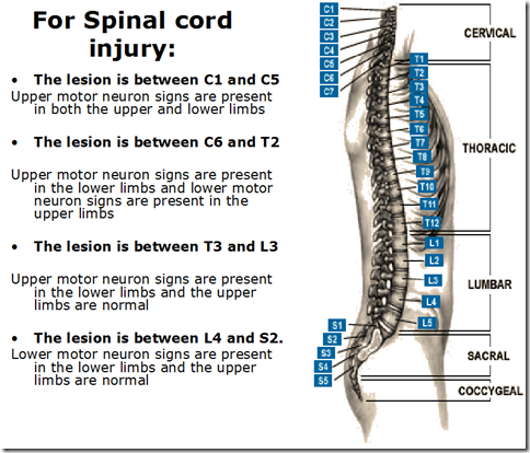
 The internal carotid arteries branch inside the brain into two major arteries, the anterior cerebral and middle cerebral arteries, as well as several minor arteries, the ophthalmic, posterior communicating, and anterior choroidal arteries. Disease and Diagnostic Testing Methods. diseases classification disease types different pathogenesis clinical associated features Vasc Endovasc Therapy Vol. Manage with MyKarger your orders simply and fast, save your favorite articles in your reading list, edit your newsletter profile and benefit from attractive discounts. costly healthaffairs Your neurologist works with doctors trained in brain and nervous system surgery (neurosurgeons), cardiologists, and others to ensure you get exactly the care you need. All Published work is licensed under a Creative Commons Attribution 4.0 International License Copyright 2022 All rights reserved. The catheter (a long, narrow, flexible tube) is introduced into the artery through the needle. Editorial Board
The carotid arteries and the vertebral arteries are two sets of arteries that carry blood from the heart to the brain. They conduct research and review medical histories to learn how diseases progress, how to treat them and which treatments may be most appropriate for people. You may be seen in the Heart Brain Clinic, one of the first such advanced multidisciplinary clinics in the United States. Read more about research in stroke and vascular disease. You will be sent an e-mail containing a link to reset your password. Read about Loyolas current clinical trials. Our clinician-researchers and scientists are committed to advancing the understanding of brain and blood vessel conditions (cerebrovascular diseases) and developing new treatments. neuron motor spinal cord lesions upper lower injury neurons umn lmn test nremt nursing injuries prep medatrio corticospinal Schedule an appointment today. Mayo Clinic's campus in Arizona, and the Mayo Clinic Health System sites in Eau Claire, Wisconsin, La Crosse, Wisconsin, and Mankato, Minnesota, are certified as Primary Stroke Centers by The Joint Commission. Electroencephalogram (EEG): A diagnostic procedure that involves placing small metal discs (electrodes) on a person's head to detect electrical impulses. Because the brain's blood supply is limited to only two sets of major arteries, it's critical that these arteries stay in good shape. 6 No. Mayo Clinic in Phoenix/Scottsdale, Ariz., is ranked highly performing for neurology and neurosurgery by U.S. News & World Report. Loyola Medicine provides comprehensive, multidisciplinary care for cerebrovascular diseases and disorders. As an academic medical center, Loyola Medicine is dedicated to improving future treatments by conducting research on new medications and protocols. Neurosurgeons can see the arteries and vessels in and around the brain, as well as the brain tissue itself, using these tests. Blood leaves the brain through the jugular and other veins.
The internal carotid arteries branch inside the brain into two major arteries, the anterior cerebral and middle cerebral arteries, as well as several minor arteries, the ophthalmic, posterior communicating, and anterior choroidal arteries. Disease and Diagnostic Testing Methods. diseases classification disease types different pathogenesis clinical associated features Vasc Endovasc Therapy Vol. Manage with MyKarger your orders simply and fast, save your favorite articles in your reading list, edit your newsletter profile and benefit from attractive discounts. costly healthaffairs Your neurologist works with doctors trained in brain and nervous system surgery (neurosurgeons), cardiologists, and others to ensure you get exactly the care you need. All Published work is licensed under a Creative Commons Attribution 4.0 International License Copyright 2022 All rights reserved. The catheter (a long, narrow, flexible tube) is introduced into the artery through the needle. Editorial Board
The carotid arteries and the vertebral arteries are two sets of arteries that carry blood from the heart to the brain. They conduct research and review medical histories to learn how diseases progress, how to treat them and which treatments may be most appropriate for people. You may be seen in the Heart Brain Clinic, one of the first such advanced multidisciplinary clinics in the United States. Read more about research in stroke and vascular disease. You will be sent an e-mail containing a link to reset your password. Read about Loyolas current clinical trials. Our clinician-researchers and scientists are committed to advancing the understanding of brain and blood vessel conditions (cerebrovascular diseases) and developing new treatments. neuron motor spinal cord lesions upper lower injury neurons umn lmn test nremt nursing injuries prep medatrio corticospinal Schedule an appointment today. Mayo Clinic's campus in Arizona, and the Mayo Clinic Health System sites in Eau Claire, Wisconsin, La Crosse, Wisconsin, and Mankato, Minnesota, are certified as Primary Stroke Centers by The Joint Commission. Electroencephalogram (EEG): A diagnostic procedure that involves placing small metal discs (electrodes) on a person's head to detect electrical impulses. Because the brain's blood supply is limited to only two sets of major arteries, it's critical that these arteries stay in good shape. 6 No. Mayo Clinic in Phoenix/Scottsdale, Ariz., is ranked highly performing for neurology and neurosurgery by U.S. News & World Report. Loyola Medicine provides comprehensive, multidisciplinary care for cerebrovascular diseases and disorders. As an academic medical center, Loyola Medicine is dedicated to improving future treatments by conducting research on new medications and protocols. Neurosurgeons can see the arteries and vessels in and around the brain, as well as the brain tissue itself, using these tests. Blood leaves the brain through the jugular and other veins.  Editor(s): Anderson, Craig S. (Sydney, NSW)
Mayo Clinic is a not-for-profit organization. Ischemia (lack of blood supply to the brain) damages brain tissue and can lead to a stroke. Because blood may easily be seen on a CT scan, it is an effective diagnostic tool for hemorrhagic strokes. 1 hospital in Minnesota, and the five-state region of Iowa, Minnesota, North Dakota, South Dakota and Wisconsin, according to U.S. News & World Report's 20222023 "Best Children's Hospitals" rankings. Our expertteam provides comprehensive care for patients to determine neurological diseases, injuries and issues interfering with critical neurological functions. It can show different types of nerve tissue as well as good images of the brain stem and posterior brain. neurology examples electrode eeg placement smartdraw Mayo Clinic's campuses in Florida and Minnesota are each certified as a Comprehensive Stroke Center by The Joint Commission, a national organization that evaluates and accredits hospitals and staff. A computer assembles the magnetic images to create an image of the arteries in the head and neck. A brain haemorrhage can produce bleeding, and this test can help detect it. The MRA can assist discover obstruction and aneurysms by showing the actual blood arteries in the neck and brain. Carotid duplex: Ultrasonography is utilised in the carotid duplex (also known as carotid ultrasound) technique to detect plaque, blood clots, or other abnormalities with blood flow in the carotid arteries. The vertebral arteries run parallel to the spine and are not visible from the outside. Stroke, carotid stenosis, vertebral stenosis, intracranial stenosis, aneurysms, and vascular abnormalities are all examples of cerebrovascular illness.
Editor(s): Anderson, Craig S. (Sydney, NSW)
Mayo Clinic is a not-for-profit organization. Ischemia (lack of blood supply to the brain) damages brain tissue and can lead to a stroke. Because blood may easily be seen on a CT scan, it is an effective diagnostic tool for hemorrhagic strokes. 1 hospital in Minnesota, and the five-state region of Iowa, Minnesota, North Dakota, South Dakota and Wisconsin, according to U.S. News & World Report's 20222023 "Best Children's Hospitals" rankings. Our expertteam provides comprehensive care for patients to determine neurological diseases, injuries and issues interfering with critical neurological functions. It can show different types of nerve tissue as well as good images of the brain stem and posterior brain. neurology examples electrode eeg placement smartdraw Mayo Clinic's campuses in Florida and Minnesota are each certified as a Comprehensive Stroke Center by The Joint Commission, a national organization that evaluates and accredits hospitals and staff. A computer assembles the magnetic images to create an image of the arteries in the head and neck. A brain haemorrhage can produce bleeding, and this test can help detect it. The MRA can assist discover obstruction and aneurysms by showing the actual blood arteries in the neck and brain. Carotid duplex: Ultrasonography is utilised in the carotid duplex (also known as carotid ultrasound) technique to detect plaque, blood clots, or other abnormalities with blood flow in the carotid arteries. The vertebral arteries run parallel to the spine and are not visible from the outside. Stroke, carotid stenosis, vertebral stenosis, intracranial stenosis, aneurysms, and vascular abnormalities are all examples of cerebrovascular illness.  People who have strokes and other brain and blood vessel conditions (cerebrovascular diseases) benefit from being evaluated and treated by the doctors of the specialty group for cerebrovascular diseases and critical care. Doppler ultrasound: On the transducer (a handheld instrument that directs high-frequency sound waves to the artery or vein being tested) and the skin over the veins of the extremity being tested, a water-soluble gel is applied. A transient ischemic attack (TIA, or mini-stroke) happens when blood flow to part of the brain is blocked or reduced for a short time. Loyolas compassionate team understands that cerebrovascular diseases can be life-changing not only for the patient but also for family members. The affected brain cells are either injured or die within minutes of being deprived of oxygen and critical nutrients. Mayo Clinic in Rochester, Minn., and Mayo Clinic in Jacksonville, Fla., are ranked among the Best Hospitals for neurology and neurosurgery by U.S. News & World Report. fazekas arteries polygon aca morphometric Tests that your Loyola doctor may order vary widely depending on your condition, and your evaluation may include: Diseases and disorders affecting the blood vessels of the brain, such as strokes, aneurysms and arteriovenous malformations (AVMs), are treated through a collaborative approach that includes neurologists, neurosurgeons, cranial base surgeons, neuroradiologists, neuro-anesthesiologists and others. Loyola takes a multidisciplinary approach to patient care and provides support services for patients and families. JavaScript is currently disabled, this site works much better if you enable JavaScript in your browser. These arteries give blood to the brain's front two-thirds. Cerebrovascular conditions treated at Loyola include: Cerebrovascular diseases may be associated with different vascular risk factors, including high blood pressure, high cholesterol, diabetes or smoking.